Jan’s Corner

Jan Hinsch died peacefully on December 31, 2023. His beautiful work and dedication to NYMS will never be forgotten.
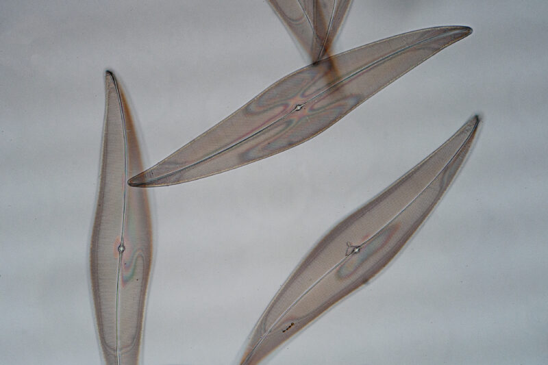

In a well aligned microscope P.a. appears to be made up of tiny, evenly spaced, black dots. Their shortest distance along the direction of the striae is 0.63 microns. If the dots can be recognized as separate entities, we call them “resolved”. If we defocus the black dots just a little in either direction they will turn into grayish-white dots. If this transition occurs smoothly upon repeated back and forth focus motion, that microscope passes inspection has having a good fine focus mechanism.
There is yet another surprise which is a colorful pattern of two vaguely insect wing-shaped zones. They are due to an airgap that exists between the underside of the cover glass and the upper surface of the diatom. The light that is reflected between the two surfaces is coherent, coming from the same light source and produce interference
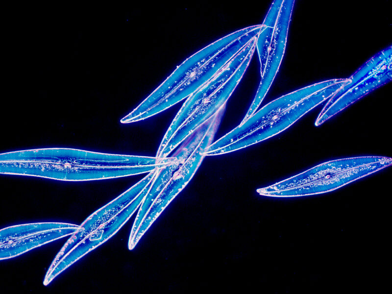
3) This time P.a. has turned a brilliant blue. The illuminating lens, the condenser, is at work. It floods the diatom not with the usual solid cone of light but with a hollow one instead. The inner angle is chosen in such a way that its light just bypasses the objective. there would be darkness were it not for the outer part of the light cone. In fact even the light that issues from the outer cone does that under such a steep angle that it too would miss the objective lens altogether were it not for the collision with the diatom. The diatom on account of its periodic structure is a form of optical grating which changes the path of light rays according to wavelenth. Red, the long waves, the least. blue the short waves the most with green in the middle. The angle then is a function of the fineness of the grating. Just by by the presence of the blue we get a good idea of the spacing of the dots. We can also see white.
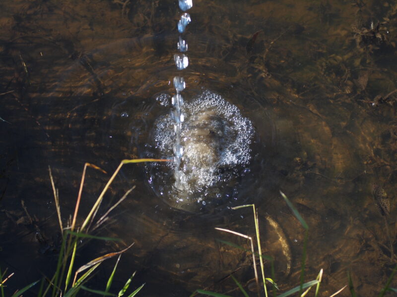
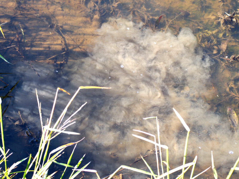

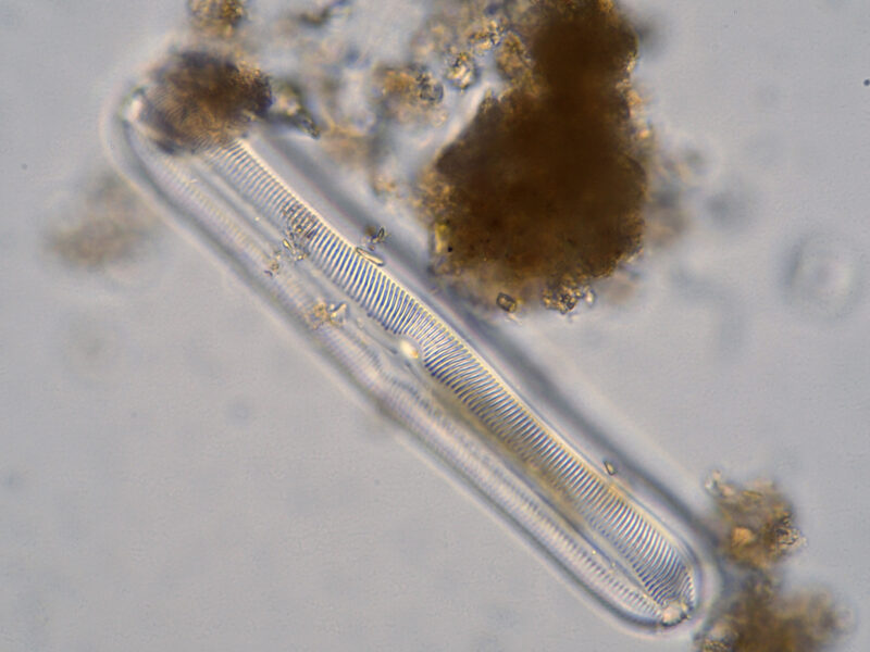
4-7) These photos were taken on a vacation trip to a New Hampshire campground. It offered a fine opportunity to check out a method of collecting diatoms that I learned about in an article by one of the members of the Quekett Microscope Club in London. Fill a glass jar with pond water pour it into a shallow pond and capture from the clouds of rising sediment a quantity of pond water. A drop of it is likely to contain several diatoms. Just one of them is shown on the micrograph, a nice reminder of a vacation trip with Wiebke, my wife.
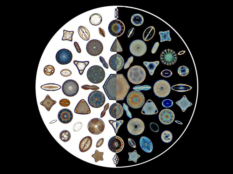
8) This micrograph shows one half of a circular arrangement of diatoms and its mirror image. Every diatom on the left (imaged in brightfield) has a symmetrical counterpart on the right (imaged in darkfield).
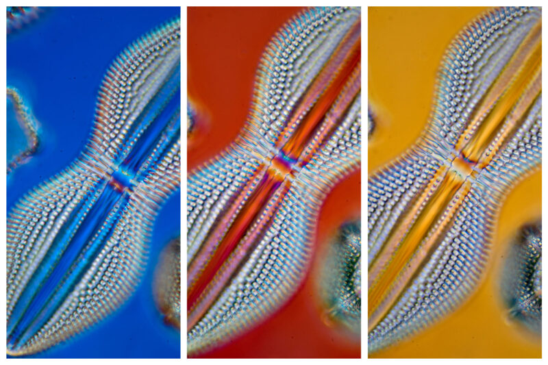
9) Three micrographs of one diatom taken in Differential Interference Contrast (DIC). This illumination method produces contrast in translucent objects, such as diatoms. It is sensitive to local changes of refraction and employs polarized light which, when combined with a crystal plate, produces the brilliant colors seen here.
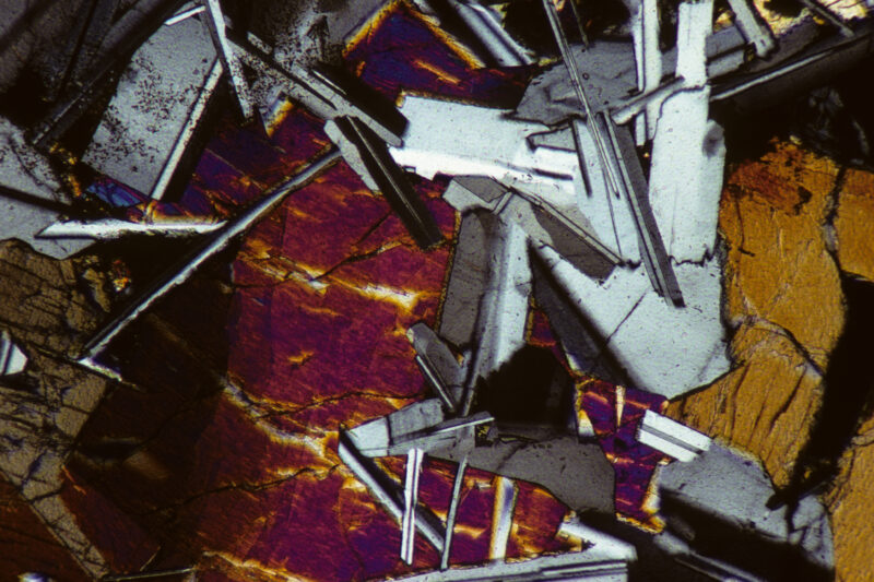
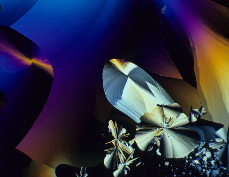
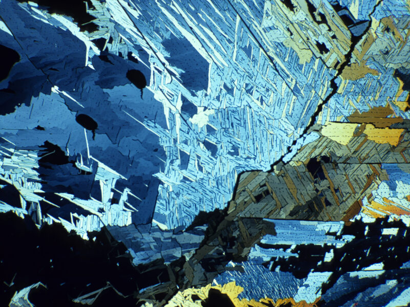
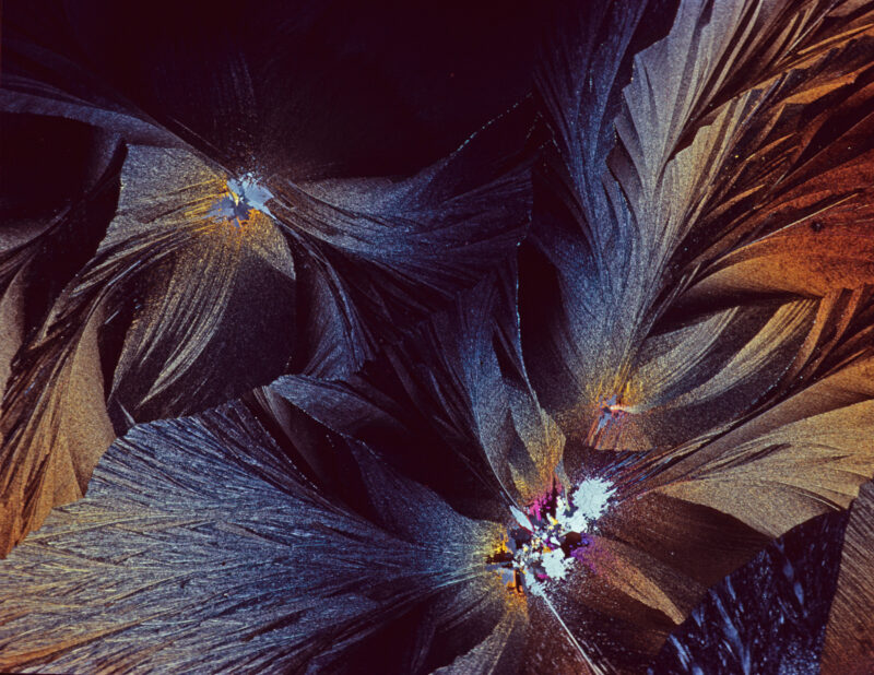
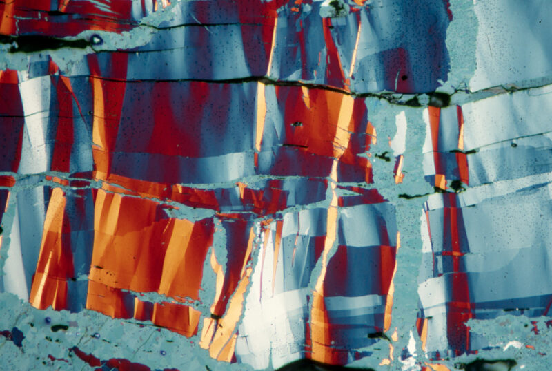
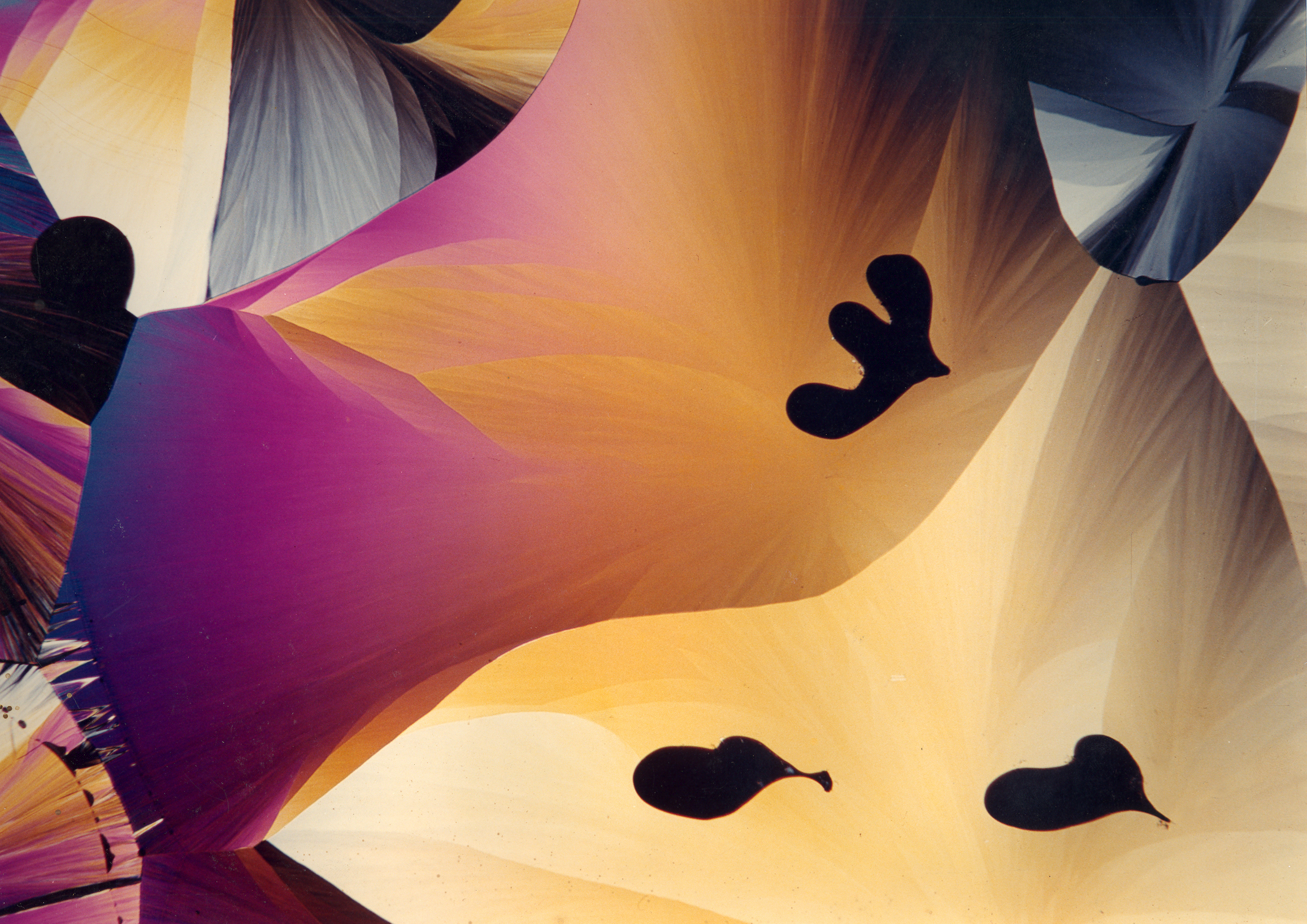
Jan’s Crystals
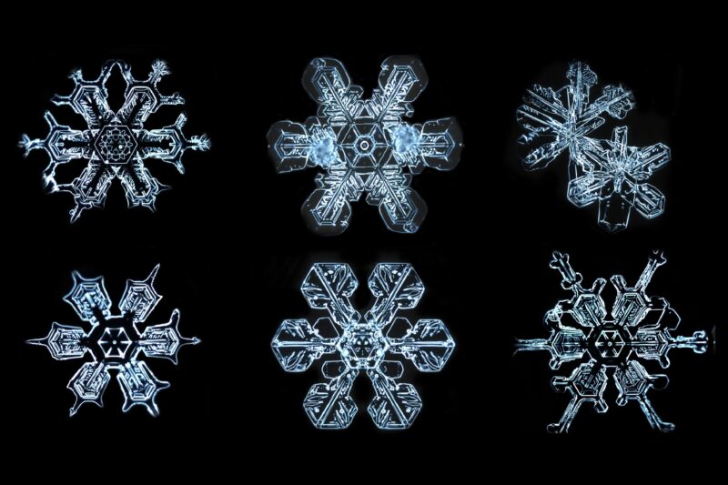
Six snowflakes
Unknown crystal
Phloroglucin
Phloroglucin
Sulfer
Covellin, Polished Ore
Unidentified crystal